Tag: SIFK

  • How Is a Medial Meniscus Posterior Root Repair Performed?

    When patients are told that a medial meniscus posterior root tear should be repaired, the next question is usually straightforward: what actually happens during the operation?
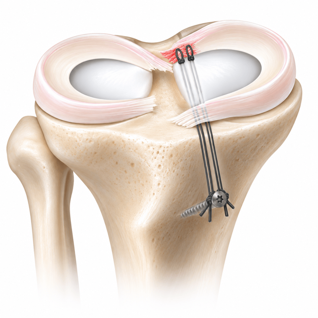
    The concept is simple. The torn posterior root is brought back to the area of the tibia where it normally attaches and is secured there so that it can heal. The execution is less simple. The root sits at the back of a tight medial compartment, and the remaining meniscal tissue may already be fragile. Good visualization, accurate tunnel or anchor placement, careful suture passage and stable fixation all matter.

    I perform the procedure arthroscopically. Standard anterior portals are used, but additional viewing access may be needed to see the posterior root directly. In selected knees, a posteromedial portal and a 70-degree arthroscope help define the native footprint more clearly. If the medial compartment is exceptionally tight, limited MCL pie-crusting can be used to improve working space rather than forcing instruments against the articular cartilage.

    Once the footprint has been identified, it is prepared and the repair construct is created. In my usual transtibial technique, a guide pin is placed at the intended root attachment and a small tibial tunnel is reamed. High-strength sutures are then passed through the meniscal root. I prefer to create two cinch-type grasping constructs with as few tissue penetrations as possible.

    That point deserves emphasis. The goal is not simply to place more sutures. Repeatedly passing a needle through degenerative meniscal tissue can damage the tissue that must ultimately hold the repair. I therefore try to plan the passes carefully and obtain a secure grasp with minimal additional trauma.

    The suture limbs are brought through the tibial tunnel to the front of the tibia and tensioned while the root is reduced to bone. Several fixation methods are available. Cortical buttons are widely used. In my practice, I generally use a cortical screw and washer as a post and tie the two suture constructs sequentially. I like the ability to control the final tension while directly watching the root dock against the tibial footprint.

    A different approach is to place a suture anchor close to the root and repair the meniscus without a long tibial tunnel. Recent biomechanical literature has generally favored suture-anchor constructs for stiffness and resistance to cyclic displacement. That does not yet mean that anchors are clinically superior in every patient. Comparative clinical studies, including recent data, have shown meaningful improvement with both anchor and transtibial techniques without a clear clinical winner.

    The choice becomes particularly interesting when root repair is combined with high tibial osteotomy. A root tunnel and the screws from an osteotomy plate can intersect. This is not only a theoretical concern; imaging-based surgical simulation studies have demonstrated that the risk depends on tunnel exit and plate position. When I combine the procedures, I plan the tunnel and plate together and sequence the operation to minimize that conflict.

    This is why medial meniscus root repair has a learning curve. The incisions are small, but the work inside the knee is precise. The surgeon has to create adequate working space, identify the correct footprint, protect the remaining meniscus, avoid unnecessary cartilage injury and maintain a stable reduction.

    Technique, however, is only one part of the result. A technically well-executed repair cannot reverse advanced osteoarthritis or poor tissue biology. I first decide whether a patient is a reasonable candidate based on cartilage status, alignment, meniscal tissue quality, activity level and the ability to follow postoperative restrictions. If repair is appropriate, then precision in the operating room becomes the next priority.

    For patients, the most useful way to think about root repair is not that the meniscus is simply ‘stitched.’ The operation is an attempt to restore the load-bearing root attachment as closely as possible while giving the tissue a realistic chance to heal.

    References

    Meng C, et al. Suture Anchor Technique Shows Superior Biomechanical Properties Compared With Transtibial Pull-Out for Posterior Medial Meniscus Root Repairs: A Systematic Review. Arthroscopy. 2026;42(7):1266-1281. PMID 41946450. DOI 10.1002/arj.70155.

    Saengpetch N, et al. Radiologic and Clinical Analysis of Posterior Medial Meniscal Root Tears: A Comparative Study of All-Suture Anchor and Transtibial Pullout Techniques. Orthop J Sports Med. 2026;14(7):23259671261464696. PMID 42534383. DOI 10.1177/23259671261464696.

    Kim JH, et al. Arthroscopic suture anchor repair versus pullout suture repair in posterior root tear of the medial meniscus: a prospective comparison study. Arthroscopy. 2011;27(12):1644-1653. PMID 21982389. DOI 10.1016/j.arthro.2011.06.033.

    Robinson JR, et al. Knotless Anchor Fixation for Transosseous Meniscal Root Repair Using Suture Tape Is Inferior Compared With Button or Screw Fixation: A Biomechanical Study. Orthop J Sports Med. 2020;8(4):2325967120912185. PMID 32341928. DOI 10.1177/2325967120912185.

    Krych AJ, et al. A simple cinch is superior to a locking loop for meniscus root repair: a human biomechanical comparison of suture constructs in a transtibial pull-out model. Knee Surg Sports Traumatol Arthrosc. 2018;26(8):2239-2244. PMID 28748490. DOI 10.1007/s00167-017-4652-1.

    LaPrade RF, et al. Cyclic displacement after meniscal root repair fixation: a human biomechanical evaluation. Am J Sports Med. 2015;43(4):892-898. PMID 25556220. DOI 10.1177/0363546514562554.

    Chang TY, et al. Biomechanical analysis of four different meniscus suturing techniques for posterior meniscal root pull-out repair: A human cadaveric study. J Exp Orthop. 2024;11(3):e70020. PMID 39318713. DOI 10.1002/jeo2.70020.

    Nejima S, et al. Risk of interference between the tibial tunnel and locking screws in medial meniscus posterior root repair and open wedge high tibial osteotomy. J Exp Orthop. 2022;9(1):25. PMID 35292866. DOI 10.1186/s40634-022-00464-0.

    Steineman BD, et al. Nonanatomic Placement of Posteromedial Meniscal Root Repairs: A Finite Element Study. J Biomech Eng. 2020;142(8):081004. PMID 31901167. DOI 10.1115/1.4045893.

    Kotipalli S, et al. Pie-crusting the medial collateral ligament is a safe and effective technique for improving visualisation and access in arthroscopic meniscal surgery: A systematic review. Knee Surg Sports Traumatol Arthrosc. 2026;34(9):3298-3313. PMID 41144741. DOI 10.1002/ksa.70103.

  • 반월상연골판 뿌리파열의 자연경과와 치료 선택

    반월상연골판 뿌리파열을 진단받으면 환자들이 가장 많이 묻는 질문이 있습니다. “수술을 꼭 해야 하나요? 그냥 지켜보면 안 되나요?”

    이 질문에 한 문장으로 답하기는 어렵습니다. 실제로 뿌리파열이 있어도 통증이 가라앉고 꽤 오랫동안 지내는 분들이 있습니다. 반대로 처음에는 비슷해 보였는데 몇 달, 몇 년 사이 관절염이 빠르게 진행하는 분도 있습니다. 문제는 처음 진단했을 때 누가 어느 쪽으로 갈지 완벽하게 골라내기 어렵다는 점입니다.

    그래서 저는 ‘지금 얼마나 아픈가’만 보지 않습니다. 이 환자가 앞으로 잃을 것이 얼마나 많은지를 같이 봅니다.

    50대처럼 비교적 젊고, 기존 관절염이 심하지 않은 무릎이라면 아직 자기 무릎으로 살아갈 시간이 많이 남아 있습니다. 이런 경우에는 뿌리봉합술로 연골판 기능을 되살릴 기회를 적극적으로 고려합니다. 수술한다고 100% 성공하는 것은 아닙니다. 재파열이나 불완전한 치유도 생길 수 있습니다. 그 가능성까지 수술 전에 설명합니다. 그래도 관절염이 심하지 않은 젊은 환자라면 관절을 보존할 수 있는 기회를 처음부터 포기하기에는 아쉬운 경우가 많습니다.

    반대로 60대 후반 이후이고 이미 관절염이 상당히 진행되어 있다면 판단이 달라질 수 있습니다. 당장 큰 수술을 하기보다 약, 주사, 활동 조절로 지내면서 경과를 보고, 결국 관절염이 더 진행하면 그때 인공관절수술로 넘어가는 전략도 현실적입니다. 나이 하나로 결정하는 것은 아닙니다. 60대 후반이라도 관절염이 거의 없고 활동성이 높다면 뿌리봉합술을 적극적으로 생각할 수 있습니다.

    현실적인 문제도 중요합니다. 뿌리봉합술 후에는 일정 기간 체중부하와 활동을 제한해야 합니다. 일을 몇 달씩 쉴 수 없는 분도 있습니다. 그런 분에게 “MRI가 이러니 무조건 수술해야 합니다”라고 말하는 것은 현실적인 진료가 아닙니다. 당장 수술이 어렵다면 우선 통증을 조절하면서 2~3개월 정도 경과를 볼 수 있습니다. 그 사이 통증이 상당히 가라앉아 생활이 가능해지는 분도 있습니다. 반대로 시간이 지나도 걷고 일하는 것이 계속 힘들다면 수술을 다시 진지하게 고려해야 합니다.

    장기 연구를 보면 조심해야 할 이유도 분명합니다. 퇴행성 내측 반월상연골판 후방 뿌리파열을 비수술로 치료한 한 장기 추적 연구에서는 평균 14년 뒤 절반 이상이 인공관절수술까지 진행했습니다. 다만 이것을 “당신도 50% 이상 인공관절을 하게 된다”는 뜻으로 받아들이면 안 됩니다. 연구 대상과 개인의 관절염 정도, 정렬, 체중, 활동도는 모두 다르기 때문입니다.

    예전에는 찢어진 연골판 일부를 잘라내는 부분절제술도 많이 했습니다. 하지만 뿌리파열에서 연골판을 잘라내는 것은 이미 망가진 ‘뿌리 기능’을 복구하지 못합니다. 최근 연구에서는 부분절제술이 관절염 진행을 막는 데 큰 도움이 되지 않았고, 적절히 선택된 환자에서는 뿌리봉합술이 부분절제술보다 장기적인 관절 보존 결과가 더 좋았습니다.

    결국 이 질환에서 중요한 것은 정답 하나를 외우는 것이 아닙니다. 환자와 보호자가 질환의 자연경과와 각 치료의 한계를 충분히 이해하고, 나이, 관절염, 다리 정렬, 활동도, 직업과 재활 여건까지 함께 놓고 선택해야 합니다.

    그리고 처음 선택이 예상과 다른 결과를 보였다고 해서 끝은 아닙니다. 그때의 무릎 상태를 다시 평가하고, 그 상황에 맞는 두 번째 선택을 하면 됩니다. 저는 반월상연골판 뿌리파열을 치료할 때 이 ‘과정을 함께 결정하는 것’이 수술 방법만큼 중요하다고 생각합니다.

    References

    Krych AJ, et al. Nonoperative Management of Degenerative Medial Meniscus Posterior Root Tears: Poor Outcomes at a Minimum 10-Year Follow-up. Am J Sports Med. 2023;51(10):2603-2607. PMID 37434486.
    Bernard CD, et al. Medial Meniscus Posterior Root Tear Treatment: A Matched Cohort Comparison of Nonoperative Management, Partial Meniscectomy, and Repair. Am J Sports Med. 2020;48(1):128-132. PMID 31765234.
    Ro KH, et al. Clinical and Radiological Outcomes of Meniscal Repair Versus Partial Meniscectomy for Medial Meniscus Root Tears: A Systematic Review and Meta-analysis. Orthop J Sports Med. 2020;8(11):2325967120962078. PMID 33241058.
    Krivicich LM, et al. Comparison of Long-term Radiographic Outcomes and Rate and Time for Conversion to Total Knee Arthroplasty Between Repair and Meniscectomy for Medial Meniscus Posterior Root Tears: A Systematic Review and Meta-analysis. Am J Sports Med. 2022;50(7):2023-2031. PMID 34251898.
    Medial Meniscus Root Repair Is Associated With Superior Outcomes As Compared With Partial Medial Meniscectomy at Minimum 5 Years: A Systematic Review and Meta-analysis. 2026. PMID 42311344.
    Surgical Timing and Outcomes in Medial Meniscus Posterior Root Tear Repair: A Systematic Review. Am J Sports Med. 2026;54(10):2560-2570. PMID 41923442.

  • Meniscus Root Tear: Do You Always Need Surgery?

    How I think about medial meniscus posterior root tears in middle-aged and older adults

    A common story is a patient in their 50s or 60s who had been doing reasonably well, then suddenly developed significant medial or posteromedial knee pain during an ordinary movement: hurrying across a street, stepping off a bus, or getting out of a car. The X-ray may show only mild arthritis. In that situation, one diagnosis I actively consider is a medial meniscus posterior root tear (MMPRT).

    Why the “root” matters

    I often explain the meniscus root using a tree analogy. A tree needs its roots firmly anchored in the ground. In the same way, the meniscus needs a secure attachment to bone to maintain tension and distribute load. When the posterior root is torn, the meniscus can extrude outward under weight bearing. The contact area decreases and focal contact pressure rises. Biomechanically, a complete root tear can behave much like loss of the functional meniscus [1].

    Diagnosis: X-rays guide treatment, MRI confirms the tear

    Weight-bearing radiographs are essential because they show the baseline osteoarthritis and alignment that will influence treatment. They do not, however, confirm a root tear. MRI is the key test for the tear itself and also shows meniscal extrusion, cartilage status, marrow edema, and subchondral insufficiency fracture of the knee (SIFK).

    What happens if you do not have surgery?

    This is the question patients ask most often. Symptoms and structural prognosis should be separated. Some patients become much more comfortable after a period of activity modification, medication, physical therapy, or injections. In my clinical practice, the acute pain can settle substantially over several months. But pain improvement does not mean that normal root function has returned.

    Long-term observational data are concerning. In a Mayo Clinic cohort of degenerative MMPRTs treated nonoperatively, 87% met failure criteria by approximately 5 years and 31% had converted to total knee arthroplasty. At a minimum 10-year follow-up, 37 of 39 living patients (95%) met the study definition of failure [2,3]. These numbers should not be presented as inevitable for every patient, but they are strong reasons not to dismiss an MMPRT simply because pain becomes tolerable.

    Who is a reasonable candidate for root repair?

    Root repair is most attractive when the knee is still worth preserving: limited osteoarthritis, acceptable cartilage status, repairable meniscal tissue, and an alignment that does not overload the medial compartment. Systematic reviews generally favor repair over partial meniscectomy or nonoperative treatment for appropriately selected patients, with less radiographic progression and lower conversion to arthroplasty [4,5].

    Age is a factor, not a stand-alone cutoff

    I do not use chronological age alone as the decision. Recent cohorts show that appropriately selected patients older than 60 can improve after medial meniscus posterior root repair, with outcomes that are not necessarily inferior to younger patients [6,7]. Evidence specifically in patients in their 70s remains less robust, so the discussion should include uncertainty, rehabilitation burden, and the possibility of later arthroplasty.

    Alignment can change the operation

    Varus alignment increases medial compartment loading. In a younger or joint-preservation candidate with meaningful varus, high tibial osteotomy (HTO) may be considered with or without root repair. Recent systematic reviews suggest possible objective advantages from combining repair with HTO, but the incremental short-term clinical benefit is not completely settled [8].

    When arthroplasty may be the better answer

    If there is advanced cartilage loss, high-grade SIFK or collapse, or established compartmental osteoarthritis, simply repairing the root may be unlikely to solve the problem. In a properly selected isolated medial-compartment pattern, unicompartmental knee arthroplasty can be considered; with more widespread advanced osteoarthritis, total knee arthroplasty may be more appropriate. The key is not to force a “meniscus-preserving” operation onto a knee that has already progressed beyond that stage.

    My decision sequence

    Confirm the root tear and assess cartilage, extrusion, and SIFK on MRI.

    Assess weight-bearing osteoarthritis and full-length limb alignment.

    Decide whether the knee remains a realistic joint-preservation candidate.

    If yes, choose between isolated root repair and alignment correction (HTO ± repair).

    Assess tissue quality, activity level, work demands, and ability to comply with protected weight bearing and rehabilitation.

    If the joint is already advanced, consider UKA or TKA according to the compartment pattern and standard arthroplasty indications.

    When the balance is uncertain, explain both pathways and make a shared decision rather than promising a guaranteed result.

    The point I emphasize to patients
    A meniscus root tear is not a diagnosis that automatically means surgery, but it is also not a tear I am comfortable dismissing. The decision is individualized because the consequences of leaving it untreated and the limitations of repair both matter.

    References

    1. Allaire R, Muriuki M, Gilbertson L, Harner CD. Biomechanical consequences of a tear of the posterior root of the medial meniscus. J Bone Joint Surg Am. 2008;90(9):1922-1931. PMID:18762653.

    2. Krych AJ, Reardon PJ, Johnson NR, et al. Non-operative management of medial meniscus posterior horn root tears is associated with worsening arthritis and poor clinical outcome at 5-year follow-up. Knee Surg Sports Traumatol Arthrosc. 2017;25(2):383-389. doi:10.1007/s00167-016-4359-8.

    3. Krych AJ, Lamba A, Wang AS, et al. Nonoperative Management of Degenerative Medial Meniscus Posterior Root Tears: Poor Outcomes at a Minimum 10-Year Follow-up. Am J Sports Med. 2023;51(10):2603-2607. doi:10.1177/03635465231185132.

    4. Lee DR, Lu Y, Reinholz AK, et al. Root Repair Has Superior Radiological and Clinical Outcomes Than Partial Meniscectomy and Nonoperative Treatment in the Management of Meniscus Root Tears: A Systematic Review. Arthroscopy. 2025;41(2):390-417. doi:10.1016/j.arthro.2024.02.017.

    5. Krivicich LM, Kunze KN, Parvaresh KC, et al. Comparison of Long-term Radiographic Outcomes and Rate and Time for Conversion to Total Knee Arthroplasty Between Repair and Meniscectomy for Medial Meniscus Posterior Root Tears: A Systematic Review and Meta-analysis. Am J Sports Med. 2022. PMID:34251898.

    6. Jackson GR, et al. Outcomes After Isolated Medial Meniscus Posterior Root Repairs Using an Anatomic Transtibial Pullout Technique in Patients Older Than 60 Years: A Matched Cohort Study. Orthop J Sports Med. 2025. PMC11905030.

    7. Husen M, Kennedy NI, Till S, et al. Benefits of Meniscal Repair in Selected Patients Aged 60 Years and Older. Orthop J Sports Med. 2022;10(9). doi:10.1177/23259671221117491.

    8. Vosoughi F, Vahedi P, Nakhjiri MT, et al. High tibial osteotomy and concurrent medial meniscus root repair provides improved objective outcomes compared to high tibial osteotomy alone for knee osteoarthritis: A systematic review. Knee Surg Sports Traumatol Arthrosc. 2025;33(9):3361-3374. doi:10.1002/ksa.12796.