When patients are told that a medial meniscus posterior root tear should be repaired, the next question is usually straightforward: what actually happens during the operation?
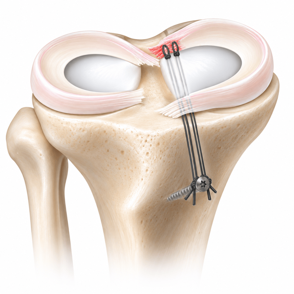
The concept is simple. The torn posterior root is brought back to the area of the tibia where it normally attaches and is secured there so that it can heal. The execution is less simple. The root sits at the back of a tight medial compartment, and the remaining meniscal tissue may already be fragile. Good visualization, accurate tunnel or anchor placement, careful suture passage and stable fixation all matter.
I perform the procedure arthroscopically. Standard anterior portals are used, but additional viewing access may be needed to see the posterior root directly. In selected knees, a posteromedial portal and a 70-degree arthroscope help define the native footprint more clearly. If the medial compartment is exceptionally tight, limited MCL pie-crusting can be used to improve working space rather than forcing instruments against the articular cartilage.
Once the footprint has been identified, it is prepared and the repair construct is created. In my usual transtibial technique, a guide pin is placed at the intended root attachment and a small tibial tunnel is reamed. High-strength sutures are then passed through the meniscal root. I prefer to create two cinch-type grasping constructs with as few tissue penetrations as possible.
That point deserves emphasis. The goal is not simply to place more sutures. Repeatedly passing a needle through degenerative meniscal tissue can damage the tissue that must ultimately hold the repair. I therefore try to plan the passes carefully and obtain a secure grasp with minimal additional trauma.
The suture limbs are brought through the tibial tunnel to the front of the tibia and tensioned while the root is reduced to bone. Several fixation methods are available. Cortical buttons are widely used. In my practice, I generally use a cortical screw and washer as a post and tie the two suture constructs sequentially. I like the ability to control the final tension while directly watching the root dock against the tibial footprint.
A different approach is to place a suture anchor close to the root and repair the meniscus without a long tibial tunnel. Recent biomechanical literature has generally favored suture-anchor constructs for stiffness and resistance to cyclic displacement. That does not yet mean that anchors are clinically superior in every patient. Comparative clinical studies, including recent data, have shown meaningful improvement with both anchor and transtibial techniques without a clear clinical winner.
The choice becomes particularly interesting when root repair is combined with high tibial osteotomy. A root tunnel and the screws from an osteotomy plate can intersect. This is not only a theoretical concern; imaging-based surgical simulation studies have demonstrated that the risk depends on tunnel exit and plate position. When I combine the procedures, I plan the tunnel and plate together and sequence the operation to minimize that conflict.
This is why medial meniscus root repair has a learning curve. The incisions are small, but the work inside the knee is precise. The surgeon has to create adequate working space, identify the correct footprint, protect the remaining meniscus, avoid unnecessary cartilage injury and maintain a stable reduction.
Technique, however, is only one part of the result. A technically well-executed repair cannot reverse advanced osteoarthritis or poor tissue biology. I first decide whether a patient is a reasonable candidate based on cartilage status, alignment, meniscal tissue quality, activity level and the ability to follow postoperative restrictions. If repair is appropriate, then precision in the operating room becomes the next priority.
For patients, the most useful way to think about root repair is not that the meniscus is simply ‘stitched.’ The operation is an attempt to restore the load-bearing root attachment as closely as possible while giving the tissue a realistic chance to heal.
References
Meng C, et al. Suture Anchor Technique Shows Superior Biomechanical Properties Compared With Transtibial Pull-Out for Posterior Medial Meniscus Root Repairs: A Systematic Review. Arthroscopy. 2026;42(7):1266-1281. PMID 41946450. DOI 10.1002/arj.70155.
Saengpetch N, et al. Radiologic and Clinical Analysis of Posterior Medial Meniscal Root Tears: A Comparative Study of All-Suture Anchor and Transtibial Pullout Techniques. Orthop J Sports Med. 2026;14(7):23259671261464696. PMID 42534383. DOI 10.1177/23259671261464696.
Kim JH, et al. Arthroscopic suture anchor repair versus pullout suture repair in posterior root tear of the medial meniscus: a prospective comparison study. Arthroscopy. 2011;27(12):1644-1653. PMID 21982389. DOI 10.1016/j.arthro.2011.06.033.
Robinson JR, et al. Knotless Anchor Fixation for Transosseous Meniscal Root Repair Using Suture Tape Is Inferior Compared With Button or Screw Fixation: A Biomechanical Study. Orthop J Sports Med. 2020;8(4):2325967120912185. PMID 32341928. DOI 10.1177/2325967120912185.
Krych AJ, et al. A simple cinch is superior to a locking loop for meniscus root repair: a human biomechanical comparison of suture constructs in a transtibial pull-out model. Knee Surg Sports Traumatol Arthrosc. 2018;26(8):2239-2244. PMID 28748490. DOI 10.1007/s00167-017-4652-1.
LaPrade RF, et al. Cyclic displacement after meniscal root repair fixation: a human biomechanical evaluation. Am J Sports Med. 2015;43(4):892-898. PMID 25556220. DOI 10.1177/0363546514562554.
Chang TY, et al. Biomechanical analysis of four different meniscus suturing techniques for posterior meniscal root pull-out repair: A human cadaveric study. J Exp Orthop. 2024;11(3):e70020. PMID 39318713. DOI 10.1002/jeo2.70020.
Nejima S, et al. Risk of interference between the tibial tunnel and locking screws in medial meniscus posterior root repair and open wedge high tibial osteotomy. J Exp Orthop. 2022;9(1):25. PMID 35292866. DOI 10.1186/s40634-022-00464-0.
Steineman BD, et al. Nonanatomic Placement of Posteromedial Meniscal Root Repairs: A Finite Element Study. J Biomech Eng. 2020;142(8):081004. PMID 31901167. DOI 10.1115/1.4045893.
Kotipalli S, et al. Pie-crusting the medial collateral ligament is a safe and effective technique for improving visualisation and access in arthroscopic meniscal surgery: A systematic review. Knee Surg Sports Traumatol Arthrosc. 2026;34(9):3298-3313. PMID 41144741. DOI 10.1002/ksa.70103.
 DR. NA | KNEE SURGEON
Leave a comment